Treatment of pseudarthrosis without surgery
After a bone fracture, the bone tissue is usually able to regenerate completely. Conservative treatment (immobilisation, orthosis, etc.) or an operation in which nails or plates made of titanium are inserted (osteosynthesis) can heal a broken bone completely without any functional restrictions remaining. This normally takes 4-8 weeks.
However, there are factors that negatively influence the healing process and increase the risk of delayed bone healing or the development of pseudarthrosis. These include inadequate immobilisation of the affected bone, a
mechanical stress or an incarceration of the surrounding tissue between the bone fragments. Infections as a result of an operation or a lack of blood flow to the bones can also be responsible for delaying or preventing the bone ends from growing together.
Some diseases, e.g. poorly controlled diabetes mellitus, medication such as cortisone or cytostatics (cancer drugs), as well as advanced age, also increase the risk of incomplete healing.
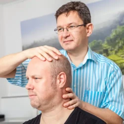
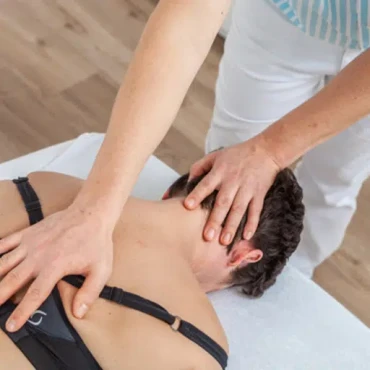
Shock wave therapy for pseudarthrosis: a gentle alternative to surgery
Numerous scientific studies have been looking at extracorporeal shock wave therapy (ESWT) as an alternative to surgical treatment of pseudarthrosis for several years. These studies confirm that ESWT has a positive influence on bone formation and leads to bone healing in up to 85% of cases, even in pseudarthrosis that has existed for years - regardless of the location and type of disease. This corresponds roughly to the same success rate of surgical procedures, but with virtually no risk or side effects! For this reason, shockwave therapy should always be the treatment of first choice in cases of delayed fracture healing or impending pseudarthrosis, as recommended by the Medical Advisory Board of the BG clinics in 2015. In our Freiburg practice for orthopaedics and osteopathy, we have been using this procedure for many years for various clinical pictures and have also seen good results in patients with pseudarthrosis.
Book an appointment now!
A quick, simple and easy way to your appointment.
What is pseudarthrosis?
What is pseudarthrosis?
The term pseudarthrosis comes from the Greek (pseudos = false, arthros = joint). It refers to a false joint formation (bony malhealing) in which the affected bone does not grow back together stably after a fracture. According to studies, delayed fracture healing or pseudarthrosis occurs in 5 to 10 % of cases. (1) According to the current definition of ESTROT (European Society of Tissue Regeneration in Orthopedics and Traumatology), the orthopaedic surgeon makes the diagnosis of pseudarthrosis if, in his opinion, a fracture does not heal without further intervention. Contrary to previous definitions, the previous duration of treatment no longer plays a role here.(2) In order to make a correct diagnosis in these cases, the orthopaedic surgeon must have a high level of knowledge and experience in this field. In addition to the medical history and a detailed physical examination, x-rays are necessary to recognise the lack of continuity of the bone. In some cases, especially if the X-ray findings are unclear, a computerised tomography (CT) scan or magnetic resonance imaging (MRI) may also be necessary.
Forms of pseudarthrosis
Orthopaedic surgeons differentiate between atrophic and hypertrophic forms of pseudarthrosis. In atrophic pseudarthrosis, the bone is no longer sufficiently supplied with blood, which means that no new bone tissue (callus) can form at the fracture site and the fracture gap cannot close. Hypertrophic pseudarthrosis - the more common form - is characterised by the formation of too much bone or cartilage tissue around the fracture site, which prevents healing. For the sake of completeness, congenital pseudarthrosis should also be mentioned, although this is very rare.
In principle, all bone fractures, regardless of whether they are treated conservatively (by immobilisation) or surgically (osteosynthesis, insertion of intramedullary nails or stable-angle plates), can lead to complications in the form of delayed fracture healing or false joint formation (pseudoarthrosis). However, certain bones are affected more frequently than others. These include, for example, the long tubular bones in the thigh (femur), shinbone (tibia) or upper arm (humerus) as well as a carpal bone, the scaphoid bone (scaphoid) and the fracture of the base of the 5th metatarsal bone (Jones fracture).
A special case is the fracture of the scaphoid bone (scaphoid fracture). The carpal bone lies in the direct neighbourhood of the radius. A scaphoid fracture is often not treated, as patients do not even consult a doctor due to the generally moderate symptoms. However, an untreated scaphoid fracture harbours a high risk of pseudarthrosis formation, which can lead to permanent discomfort, serious limitations and arthrosis in the wrist.
How does pseudarthrosis manifest itself?
Pseudarthrosis does not develop suddenly, but is subject to a gradual process. Redness and swelling over the fractured area are typical. As the stability of the bone is severely reduced in some cases, this often leads to malalignment, restricted movement, joint problems and muscle atrophy in the affected body part or limb, the function of which is then often severely restricted. Most patients are particularly distressed by the severe pain, which initially only occurs during movement, but can also occur at rest in advanced stages. Without therapy, this pain does not diminish but, on the contrary, becomes increasingly severe and therefore chronic.
Pseudarthrosis surgery is complex and associated with considerable risks
Pseudarthrosis is a long-term condition that does not heal on its own and therefore requires treatment. Surgery is still the standard treatment. This is a procedure that is often more extensive than the first operation, in which a partial resection of the affected bone is performed and bone tissue is usually transplanted from the iliac crest into the fracture gap or foreign bone or bone replacement material is used. The surgical site must then be immobilised with an external fixator or stable-angle plates. This means that numerous and not insignificant complications are possible with this procedure. In addition to bleeding, secondary bleeding and haematoma, infections, wound healing disorders, scarring and chronic post-operative pain can occur. Nerve injuries can result in sensory disturbances or paralysis. In some cases, reduced mobility or joint stiffness may occur. Sudeck's syndrome (CRPS) cannot be ruled out either. In this case, the bone is severely degraded, resulting in painful inflammation. All of these are risks of complications, which of course do not necessarily occur, but only in some cases. However, the patient is always subjected to considerable stress during the procedure. In addition, the entire post-operative follow-up treatment virtually starts all over again. This is sometimes associated with long periods of rehabilitation and absence from work.